DETECT
La détection : notre plus belle mission !
Une lésion détectée est une lésion en sursis, qui sera réséquée ou prise en charge quoi qu’il arrive. Au contraire, passer à côté d’un cancer, d’un adénome ou d'une lésion festonnée peut aboutir à des situations catastrophiques. De même, un patient non détecté n’est plus un lanceur d’alerte pour dépister une famille entière…
L’importance du Taux de Détection d’Adénome (TDA) comme indicateur de qualité de sa puissance de détection est désormais bien connu. En pratique, les cancers d’intervalle tendent à devenir rares dans la patientèle des gastroentérologues dont le TDA dépasse les 25 %[4,6-7]. Cet objectif doit devenir le strict minimum acceptable. De nombreuses techniques sont actuellement développées pour améliorer la détection chez tous les gastroentérologues, des plus expérimentés aux moins performants. Le seuil de ces 25 % doit être dépassé grâce aux différents points d’optimisation regroupés sous l’acronyme DETECT :
- La Détersion du côlon par une préparation de qualité est impérative et l’examen n’a de valeur que dans ces conditions[8].
- L’Exhaustivité de l’examen colique est le point le plus important. L’objectif est de déplisser le côlon, de voir derrière les plis, de revenir sur les zones aveugles, de répéter l’examen de certains segments… Parmi les techniques permettant d’améliorer la surface de côlon vue, les différents capuchons, qu’ils soient transparents ou à extensions latérales[9] (Endocuff vision®, Endorings®), permettent de mieux voir derrière les plis et ont démontré leur intérêt pour augmenter le TDA[10], en particulier pour les détecteurs moyens. D’autres techniques existent pour augmenter la surface de côlon examiné :
- la coloscopie en immersion sous-marine,
- la rétrovision systématique dans le côlon droit ou dans le rectum,
- les endoscopes avec plusieurs optiques mais dont les résultats sont plus discutés.
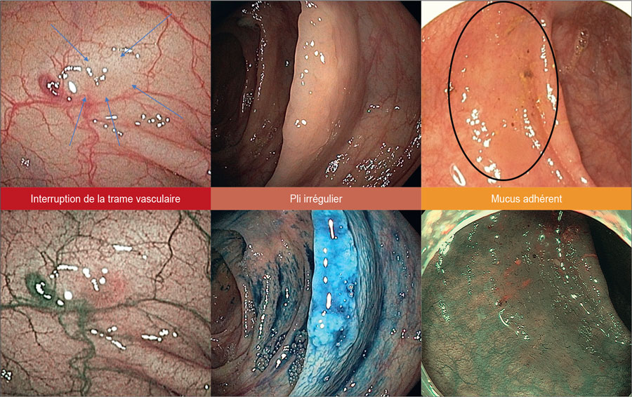
- La Traque doit être acharnée et permanente. Il ne suffit pas de voir un segment colique pour détecter une lésion : il faut dépister toute interruption de la trame vasculaire de fond, toute irrégularité de la forme d’un pli, toute zone de mucus ou de selles adhérentes dans une zone non déclive. Cette étape de traque demande motivation et minutie.
- Les Endoscopes doivent être performants, de haute-définition, avec un champ large et une chromoendoscopie virtuelle. Les nouvelles générations d’endoscopes permettent de diminuer le taux d’adénomes manqués par rapport aux générations plus anciennes[11].
- L’Entraînement doit être permanent pour maîtriser les différentes techniques : on ne détecte bien que ce que l’on connaît.
- Les séances d’entraînement de Caractérisation sur des images de lésions coliques doivent se multiplier afin de s’accoutumer à voir et revoir des lésions caractéristiques.
- Se Tester pour connaître son TDA est indispensable et entre dans cette démarche d’entraînement et de progression. Il s’agit de le faire progresser bien au-delà des 25 % recommandés, de même que le taux de détection des festonnées qui devrait dépasser les 7,5 %.

Figure 4 : Points clefs de détection.
[1] Gandilhon C, Soler-Michel P, Vecchiato L et al. A motivational phone call improves participation to screening colonoscopy for those with a positive FIT in a national screening programme (NCT 03276091). Dig Liver Dis. 2018 ; 50 : 1309-1314.
[2] Gimeno-García AZ, de la Barreda Heuser R, Reygosa C et al. Impact of a 1-day versus 3-day low-residue diet on bowel cleansing quality before colonoscopy : a randomized controlled trial. Endoscopy. 2019 ; 51 : 628-636.
[3] Mangira D, Ket S, Dwyer J et al. Augmentation with pre-emptive macrogol-based osmotic laxative does not significantly improve standard bowel preparation in unselected patients : A randomized trial. JGH Open Open Access J Gastroenterol Hepatol. 2019 ; 3 : 37-380.
[4] Kaminski MF, Thomas-Gibson S, Bugajski M et al. Performance measures for lower gastrointestinal endoscopy : a European Society of Gastrointestinal Endoscopy (ESGE) quality improvement initiative. United Eur Gastroenterol J. 2017 ; 5 : 309-334.
[5] Fabritius M, Jacques J, Gonzalez J-M et al. A simplified table mixing validated diagnostic criteria is effective to improve characterization of colorectal lesions : the CONECCT teaching. Endosc Int Open. 2019 ; 07 : E1197-E1206.
[6] Kaminski MF, Regula J, Kraszewska E et al. Quality indicators for colonoscopy and the risk of interval cancer. N Engl J Med. 2010 ; 362 : 1795-1803.
[7] Corley DA, Levin TR, Doubeni CA. Adenoma detection rate and risk of colorectal cancer and death. N Engl J Med. 2014 ; 370 : 1298-306.
[8] Clark BT, Rustagi T, Laine L. What level of bowel prep quality requires early repeat colonoscopy : systematic review and meta-analysis of the impact of preparation quality on adenoma detection rate. Am J Gastroenterol. 2014 ; 109 : 1714-1723 ; quiz 1724.
[9] Sola-Vera J, Catalá L, Uceda F et al. Cuff-assisted versus cap-assisted colonoscopy for adenoma detection : results of a randomized study. Endoscopy. 2019 ; 51 : 742-749.
[10] Williet N, Tournier Q, Vernet C et al. Effect of Endocuff-assisted colonoscopy on adenoma detection rate : meta-analysis of randomized controlled trials. Endoscopy. 2018 ; 50 : 846-860.
[11] Pioche M, Denis A, Allescher H-D et al. Impact of 2 generational improvements in colonoscopes on adenoma miss rates : results of a prospective randomized multicenter tandem study. Gastrointest Endosc. 2018 ; 88 : 107-116.
[12] Tate DJ, Awadie H, Bahin FF et al. Wide-field piecemeal cold snare polypectomy of large sessile serrated polyps without a submucosal injection is safe. Endoscopy. 2018 ; 50 : 248-252.
[13] Binmoeller KF, Weilert F, Shah J et al. „Underwater“ EMR without submucosal injection for large sessile colorectal polyps (with video). Gastrointest Endosc. 2012 ; 75 : 1086-1091.

